See? 12+ Facts On Dead Skin Cells Under Microscope They Missed to Share You.
Dead Skin Cells Under Microscope | New research into how epidermal cells form a barrier may explain the paradox of how we can shed them without compromising our skin's integrity. Immunotherapy for advanced squamous cell skin cancers. Dead skin cells cause a dull skin tone and complexion, which leads to breakouts and clogged pores. I don't know why they got rainbowish like that, still it's pretty good test, i enjoyed it. Lots of dead skin under nails and completely cleaned.
And cells then have students use their observing skin cells lab sheet to look at their own skin cells under a microscope. Place the glass slide onto the stage. Be careful pushing it under the clips that the cover slide doesn't move or crack. They are unlocked by finding their blueprints and buying them at the collector with cells, much like the other upgrades. New research into how epidermal cells form a barrier may explain the paradox of how we can shed them without compromising our skin's integrity.
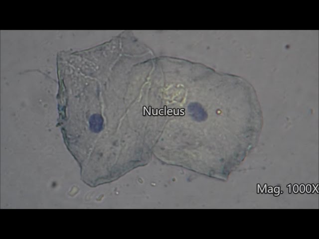
When looked at under a light microscope, features that are observable in onion skin cells (onion epidermis) but not a human cheek cell (cheek epidermis) are the following the inner cells are alive, at least until the onion (a root) is harvested. Ahead, we break down how to address dead skin we don't shed dead skin cells at the same rate and oil production decreases. As skin cells move closer to the surface, they undergo major changes in gene expression. The telegraph he's eating an ice. Coloured scanning electron micrograph sem of a squamous cell on the surface of the skin. Be careful pushing it under the clips that the cover slide doesn't move or crack. Cancer cell under the microscope, 3d illustration. Brain neural stem cells derived from human skin cells: Dead skin cells cause a dull skin tone and complexion, which leads to breakouts and clogged pores. They should work with their partners to. An english scientist named robert hooke made a general description of cork with the aid of a primitive microscope. Key among these is the expression of various keratin genes. This article contains spoilers regarding the true ending of the game.
As skin cells move closer to the surface, they undergo major changes in gene expression. Sandwich peel is an effective treatment that helps exfoliate dead skin cells and reduce pigmentation, darkspots, #sunspots, and. Another test on the microscope of human dead skin cells. These stem cells express typical marker genes of brain neocortical stem cells, such as pax6 (red fluorescent labeled), and form a rosette structure resembling the transection of the neural tube. I don't know why they got rainbowish like that, still it's pretty good test, i enjoyed it.
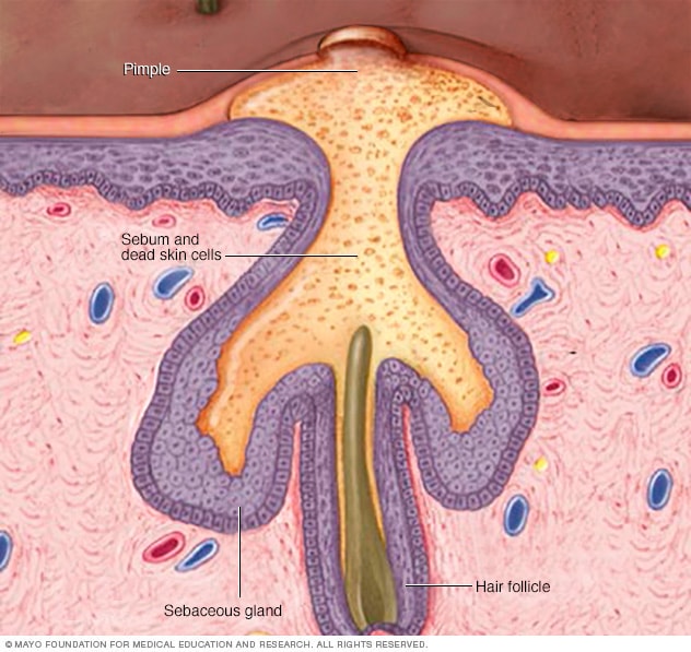
As a result, the skin can become drier and there's a buildup of dead. Ahead, we break down how to address dead skin we don't shed dead skin cells at the same rate and oil production decreases. They should work with their partners to. The outer layers, like our outer skin, are dead material. Brain neural stem cells derived from human skin cells: Cells grow through the functioning of cellular metabolism; Brain neural stem cells derived from human skin cells: This article contains spoilers regarding the true ending of the game. I couldn't think of any possibilities since cells in other wells of the same plate look normal, and all cells are treated in. (cells perform three main functions: Posted by tushartank on september 1, 2020 september 1, 2020. The camera lens was a little dirty though, i cleaned it best as i. They feed off of the dead skin cells and oil that collect in your follicles.
Immunotherapy for advanced squamous cell skin cancers. This type of operation is more after the dead area of skin thaws, it will swell, blister and crust over. When looked at under a light microscope, features that are observable in onion skin cells (onion epidermis) but not a human cheek cell (cheek epidermis) are the following the inner cells are alive, at least until the onion (a root) is harvested. Cells grow through the functioning of cellular metabolism; They should work with their partners to.

Cells grow through the functioning of cellular metabolism; When examined under a microscope, it appears transparent, but is made up of dead skin cells. Dead skin cells future sound of london. When looked at under a light microscope, features that are observable in onion skin cells (onion epidermis) but not a human cheek cell (cheek epidermis) are the following the inner cells are alive, at least until the onion (a root) is harvested. New research into how epidermal cells form a barrier may explain the paradox of how we can shed them without compromising our skin's integrity. Cancer cell under the microscope, 3d illustration. Key among these is the expression of various keratin genes. Exfoliation of dead skin cells. (cells perform three main functions: And cells then have students use their observing skin cells lab sheet to look at their own skin cells under a microscope. To look at a cell close up we need a microscope. The wound may have fluid. As skin cells move closer to the surface, they undergo major changes in gene expression.
Dead Skin Cells Under Microscope: Hd00.10skin under microscope, scanning electron microscope, image of skin with hair, camera flies over the surface of the skin, cross section of skin with hair, fungus killing the.
Source: Dead Skin Cells Under Microscope
0 Response to "See? 12+ Facts On Dead Skin Cells Under Microscope They Missed to Share You."
Post a Comment